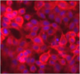
DiD is a far-red fluorescent, lipophilic carbocyanine and widely used as a lipophilic tracer. It is weakly fluorescent in water but highly fluorescent when incorporated into membranes. It has an extremely high extinction coefficient and short excited-state lifetimes (~1 nanosecond) in lipid environments. Once applied to cells, the dye diffuses laterally within the plasma membrane.
|
Specifications: |
|
|
| Excitation/Emission: | 645/665 nm | |
| Shipping Condition: | Ambient | |
| Storage Conditions: | -20ºC, protect from light | |
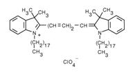
| Molecular Formula: | C61H99ClN2O4 | |
| Molecular Weight: | 959.92 | |
| CAS Number: | 127274-91-3 | |
|
||
| Protocol (PDF): | C018 |
| MSDS (PDF): | MSDS-C018 |
| COA (PDF): | C018 |
Reference:
Cytometry (2002) 48:167-176
Blood (2003) 101:594-601
Blood (2010) 115:5347-5354
Frequently Asked Questions (FAQs)
I need to look at live cell morphology deformation over the course of a few hours. What sort of membrane dye would be useful for this?
Lipophilic cyanine dyes, such as DiO (Cat. No. C016), DiI (Cat. No. C017), DiD (Cat. No. C018) or DiR (Cat. No. C019), are commonly used. The longer the alkyl chain on the dye, the better the retention in lipophilic environments.
I’m labeling live cells with DiI or DiD lipophilic cyanine dyes. DiI gives a nice even membrane labeling, but DiD is more “spotty”. What can be done?
This is expected. DiD (which is far-red fluorescent) is never as uniform as DiI (which is orange fluorescent). If uniformity is desired, try increasing the label time and concentration, but it still isn’t likely to be as uniform as DiI.
I want to perform a cell fusion assay, where one cell line is labeled with one color and the other cell line with another color, and combine with a nucleic acid stain. What do you recommend?
A typical method is to label one cell line with orange fluorescent DiI and the other cell line with green fluorescent DiO. These orange and green lipophilic cyanine dyes will stain the membranes of cells. Cells that fuse will then have both dyes, yielding a yellow color (when images are overlaid or cells are imaged in a dual-bandpass filter). These live cells can then be labeled with Hoechst 33342 (a cell-permeant blue DNA stain comparable in wavelength to DAPI), but only as an endpoint just before imaging (since DNA stains can interrupt DNA function).