Mycoplasma infections are relatively common in laboratory cell cultures; it has been estimated that between 5% and 35% of all cell cultures are infected. Mycoplasmas have been shown to alter the growth rate of cells in culture, induce chromosomal aberrations, influence amino acid and nucleic acid metabolism and cause membrane aberrations. Several methods have been developed to detect mycoplasma including direct culture in special growth media, enzyme-linked immunoassay, immunofluorescence staining, PCR, biochemical detection and fluorescent nucleic acid stains.
ABP Biosciences has developed two methods for fast and sensitive detection of Mycoplasma infection: MycoCheck™ Mycoplasma PCR Detection Kit and MycoCheck™ Mycoplasma Stain Kit.
MycoCheck™ Mycoplasma PCR Detection Kit
The MycoCheck™ Mycoplasma PCR Detection Kit allows for fast and reliable identification of mycoplasma contamination in cell cultures. Mycoplasma DNA in the cell culture supernatant is amplified via PCR and visualized using gel electrophoresis. In addition to the short detection process (less than 2 hours), the easy handling and high sensitivity makes this Mycoplasma PCR Detection Kit a convenient tool for routine examination of cell cultures and media. The kit contains an optimized PCR master mix and a positive control. The primers in the PCR master mix are highly specific to the conserved rRNA region in the mycoplasma genomes and can detect all well-known mycoplasma genera, including the commonly encountered ones in cell cultures, such as M. orale, M. hyorhinis, M. laidlawii, M. salivarium, M. arginini, M. fermentans, M. hominis, and M. pneumoniae. Mycoplasma positive samples can be easily recognized by a distinctive PCR product ranging in size from 400 to 600 bp.

Figure 1. The PCR fragments of the commonly encountered mycoplasmas in cell cultures
MycoCheck™ Mycoplasma Stain Kit
The MycoCheck™ Mycoplasma Stain Kit provides an ultrasensitive, rapid and simple fluorescence microscopic assay for the visual identification of mycoplasma infection in laboratory cell cultures. In order to detect mycoplasma, the fluorescent MycoFluor reagent is added directly to the culture medium, with or without cells, and the stained sample is then examined under a fluorescence microscope. The excitation and emission spectra of MycoFluor reagent bound to dsDNA are similar to dsDNA bound to DAPI, with excitation maxima around 350-360nm and emission maxima around 450-460nm. MycoFluor reagent can be excited either with a xenon mercury arc lamp or a UV laser and is detected through a blue filter.
Mycoplasma staining with MycoFluor reagent appears as a fine particulate or filamentous staining over the cytoplasm at 100X magnification. Nuclei of the cells are also brightly stained by this method and thereby act as endogenous positive control for the staining procedure.
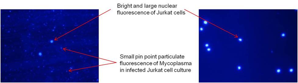
Figure 2. The Jurkat cell culture stained with MycoCheck™ Mycoplasma Stain Kit
Related Products
- Nucleic Acid Gel Stains
- Nucleic Acid Quantitation
- Labeled Nucleotides
- Protein Detection and Quantitation
- Cell Structure Probes
- Secondary Antibody and Streptavidin
- Cell Proliferation & Viability Assay
- Cell Apoptosis Assay
- Andy Fluor™ Dyes
- Ion Indicators
- Transfection Reagent
- Luciferase Assay Kit
- ECL Western Blot Reagent