Introduction
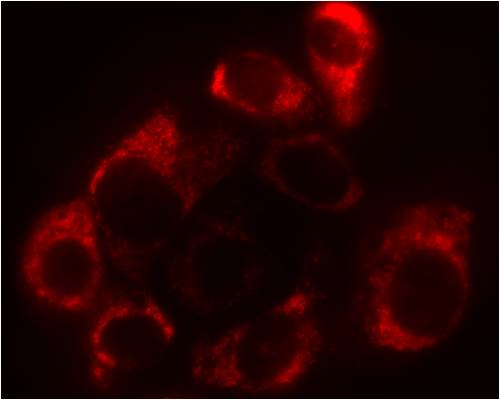
MitoTrack™ Red CMXRos (Known as MitoTracker® Red CMXRos, Trade Mark of Molecular Probes) is an red-fluorescent dye that stains mitochondria in live cells and its accumulation is dependent upon membrane potential. The dye is well-retained after aldehyde fixation.
| Specifications: |
|
|
| Excitation/Emission: | 580/600 nm | |
| Shipping Condition: | Ambient | |
| Storage Conditions: | -20ºC, protect from light | |
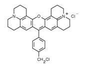
| Molecular Formula: | C32H32Cl2N2O | |
| Molecular Weight: | 531.52 | |
| CAS Number: | 167095-09-2 | |
|
||
To Order
Documents
| Protocol (PDF): | C042 |
| MSDS (PDF): | MSDS-C042 |
| COA (PDF): | C042 |
Reference:
Iuso A, Scacco S, Piccoli C, Bellomo F, Petruzzella V, Trentadue R, Minuto M, Ripoli M, Capitanio N, Zeviani M, Papa S
J Biol Chem (2006) 281:10374-10380
J Biol Chem (2006) 281:10374-10380
Nagaraj R, Gururaja-Rao S, Jones KT, Slattery M, Negre N, Braas D, Christofk H, White KP, Mann R, Banerjee U,
Genes Dev (2012) 26:2027-2037
Product usage: Proliferation, DNA Synthesis, Click-iT EdU, Imaging, in vivo
Genes Dev (2012) 26:2027-2037
Product usage: Proliferation, DNA Synthesis, Click-iT EdU, Imaging, in vivo
Zhao T, Huang X, Han L, Wang X, Cheng H, Zhao Y, Chen Q, Chen J, Cheng H, Xiao R, Zheng M,
J Biol Chem (2012) 287:23615-23625
Product usage: Lysotracker, ER-Tracker, Mitotracker, Autophagy, Imaging
J Biol Chem (2012) 287:23615-23625
Product usage: Lysotracker, ER-Tracker, Mitotracker, Autophagy, Imaging
Frequently asked questions (FAQs)
It looks like my Mitotracker™ dye is staining more than just the mitochondria. Why?
This is typically a result of using too high of a concentration of the MitoViewr™ dye. Most organic dyes are used in the low micromolar range. The MitoView™ dyes are used at a much lower concentration, around 50–200 nanomolar. Higher concentrations can cause background fluorescence and non-mitochondrial staining.